45 heart structure and labels
The Anatomy of the Heart, Its Structures, and Functions - ThoughtCo The heart is the organ that helps supply blood and oxygen to all parts of the body. It is divided by a partition (or septum) into two halves. The halves are, in turn, divided into four chambers. The heart is situated within the chest cavity and surrounded by a fluid-filled sac called the pericardium. This amazing muscle produces electrical ... Human Heart Diagram Labeled | Science Trends List Of Heart Structures Heart Chambers Ventricles - The bottom two heart chambers. Atra - The upper two heart chambers. Wall Of The Heart Sinoatrial Node - A collection of tissue that releases electrical impulses and defines the rate of contraction for the heart. Atrioventricular Bundle - The fibers which transmit cardiac impulses.
Chapter 19: The Heart Flashcards | Quizlet •Allows heart to beat without friction, gives it room to expand and resists excessive expansion •Parietal pericardium-tough outer, fibrous layer of connective tissue-inner serous layer •Visceral pericardium (a.k.a. epicardium of heart wall)-serous lining of sac turns inward at base of heart to cover the heart surface

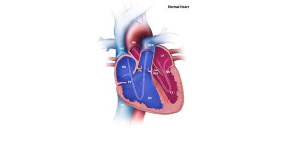
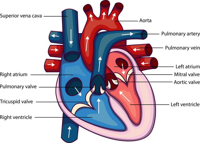
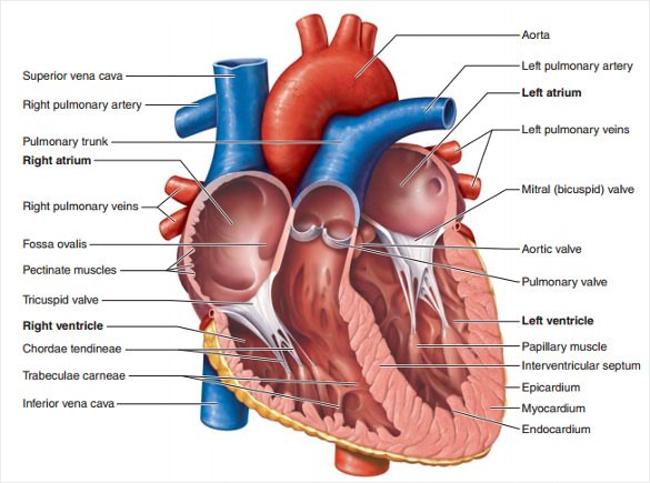
Heart structure and labels
A Diagram of the Heart and Its Functioning Explained in Detail The heart blood flow diagram (flowchart) given below will help you to understand the pathway of blood through the heart.Initial five points denotes impure or deoxygenated blood and the last five points denotes pure or oxygenated blood. 1.Different Parts of the Body ↓ 2.Major Veins ↓ 3.Right Atrium ↓ 4.Right Ventricle ↓ 5.Pulmonary Artery ↓ 6.Lungs Dapagliflozin - Wikipedia Dapagliflozin, sold under the brand names Farxiga (US) and Forxiga (EU) among others, is a medication used to treat type 2 diabetes. It is also used to treat adults with certain kinds of heart failure and chronic kidney disease.. Common side effects include hypoglycaemia (low blood sugar), urinary tract infections, genital infections, and volume depletion (reduced amount of … Eplerenone: a blood pressure medicine used to treat heart failure It’s used to treat heart failure and reduce the risk of you having other heart problems or a stroke. It also helps to stop heart failure getting worse. It can sometimes be used to treat a condition called hyperaldosteronism. This is when your body makes too much aldosterone, a hormone that controls your blood pressure. Eplerenone comes as tablets and is only available on …
Heart structure and labels. Heart (Human Anatomy): Overview, Function & Structure | Biology The heart is a muscular organ that pumps blood throughout the body. It is located in the middle cavity of the chest, between the lungs. In most people, the heart is located on the left side of the chest, beneath the breastbone. The heart is composed of smooth muscle. It has four chambers which contract in a specific order, allowing the human ... Structure of the Heart | SEER Training - National Cancer Institute The human heart is a four-chambered muscular organ, shaped and sized roughly like a man's closed fist with two-thirds of the mass to the left of midline. The heart is enclosed in a pericardial sac that is lined with the parietal layers of a serous membrane. The visceral layer of the serous membrane forms the epicardium. Layers of the Heart Wall Diagrams, quizzes and worksheets of the heart | Kenhub Labeled heart diagrams Take a look at our labeled heart diagrams (see below) to get an overview of all of the parts of the heart. Once you're feeling confident, you can test yourself using the unlabeled diagrams of the parts of the heart below. Labeled heart diagram showing the heart from anterior Unlabeled heart diagrams (free download!) Heart Anatomy: size, location, coverings and layers : Anatomy & Physiology Heart Anatomy. The heart is around the size of a fist and weighs between 250-350 grams (less than a pound). Enclosed within the mediastinum, the medial cavity of the thorax, the heart extends obliquely from the second rib to the fifth intercostal space. It rests on the superior surface of the diaphragm, lies posterior to the sternum and ...
PDF Heart Structure - Indiana The heart is an organ about the size of a fist. It is made of muscle and pumps blood through the body. Tube-like structures called blood vessels carry blood through the body and heart. The heart and blood vessels make up the cardiovascular system. Structure of the Heart The heart has four chambers: two upper chambers call Heart Diagram with Labels and Detailed Explanation - Collegedunia The heart is located under the ribcage, between the lungs and above the diaphragm. It weighs about 10.5 ounces and is cone shaped in structure. It consists of the following parts: Heart Detailed Diagram Heart - Chambers There are four chambers of the heart . The upper two chambers are the auricles and the lower two are called ventricles. Human Heart Models | Heart Anatomy Models | Vitality Medical The heart model with labels is hand-painted with vivid colors to illustrate the papillary muscles, heart valves, and adjacent structures. Sort By 4 Items Magnetic Heart Model, Life Size, 5 Parts $327.45 View Details Human Heart Model $450.66 - $566.36 View Details Classic Heart Model $81.03 View Details Magnetic Heart Model, Life Size, 5 Part G01 Fellow of the American Heart Association (FAHA) For those who qualify, election as a Fellow of the American Heart Association recognizes your scientific and professional accomplishments, volunteer leadership and service. By earning the right to include the initials FAHA among your credentials, you let colleagues and patients know that you have been welcomed into one of the world’s most eminent organizations of …
Nutritionist Pro™ | Diet Analysis, Food Label, Menu Creation ... Designed and managed by registered dietitians for your comprehensive nutrition analysis needs. From food labels to menus to recipe calculations, Nutritionist Pro™ makes all your food science needs a simple and streamlined process. Since 1982 over 1,000,000 have relied on the Nutritionist Pro™ family of products. label heart anatomy heart worksheet label parts system circulatory interactive biology liveworksheets structure pdf cardiovascular science Image Result For Vein Lab Model Labeled | Anatomy Models Labeled heart labeled anatomy models lab valve inside septum right blood physiology different side vein valves left Label Heart Anatomy Diagram Printout - EnchantedLearning.com Every day, the heart pumps about 2,000 gallons (7,600 liters) of blood, beating about 100,000 times. Label the heart anatomy diagram below using the heart glossary. Note: On the diagram, the right side of the heart appears on the left side of the picture (and vice versa) because you are looking at the heart from the front. Enchanted Learning Search System - Wikipedia Man-made systems may have such views as concept, analysis, design, implementation, deployment, structure, behavior, input data, and output data views. A system model is required to describe and represent all these views. Systems architecture A systems architecture, using one single integrated model for the description of multiple views, is a kind of system model. …
Human Heart (Anatomy): Diagram, Function, Chambers, Location in ... - WebMD Chambers of the Heart The heart is a muscular organ about the size of a fist, located just behind and slightly left of the breastbone. The heart pumps blood through the network of arteries and...
147 Heart Anatomy With Labels Premium High Res Photos - Getty Images Browse 147 heart anatomy with labels stock photos and images available, or start a new search to explore more stock photos and images. of 3. NEXT.
Structure and Function of the Heart - News-Medical.net The heart is the main organ in the circulatory system, the structure is primarily responsible for delivering blood circulation and transportation of nutrients in all parts of the body. This ...
How the Heart Works: Diagram, Anatomy, Blood Flow - MedicineNet The heart is an amazing organ. It starts beating about 22 days after conception and continuously pumps oxygenated red blood cells and nutrient-rich blood and other compounds like platelets throughout your body to sustain the life of your organs.; Its pumping power also pushes blood through organs like the lungs to remove waste products like CO2.; This fist-sized powerhouse beats (expands and ...
Eating for a healthy heart - Baker This may help to improve your overall heart health. Unhealthy ‘saturated and trans’ fats should be limited. This fact sheet will help you with: choosing healthy fats and sources; practical ways to include this into your diet; reducing unhealthy fats in your diet; how to read nutrition food labels. Download fact sheet
Human Heart - Anatomy, Functions and Facts about Heart - BYJUS To know more about the human heart structure and function, or any other related concepts such as arteries and veins, ... Practice your understanding of the heart structure. Drag and drop the correct labels to the boxes with the matching, highlighted structures. Instructions to use: Hover the mouse over one of the empty boxes. One part in the image gets highlighted. Identify the …
How would you label the structures (both external and internal) of a dissected pig's heart? - Quora
heart diagram and labels Nervous system chart anatomy human body diagram physiology study medical organs endocrine diagrams structure labeled nerve skeleton heart nursing academic. Fig 2 gross anatomy of the heart (c). Heart sheep dissection anatomy cow ventricle atrium human blood between differences pressure cut half valve practical cat edu quora labeled heart ...
called myocardium science External Structure Of Human Heart Anatomy structure of human heart ...
Heart Blood Flow | Simple Anatomy Diagram, Cardiac Circulation ... - EZmed One of the first things you will notice if you look at the 12 steps is the pattern between the right and left side of the heart is similar. Step 1 and 6 involve a blood vessel, which makes sense as this is how blood enters and exits that side of the heart. Steps 2-5 involve a chamber, valve, chamber, and valve.
Label the heart — Science Learning Hub Label the heart Interactive Add to collection In this interactive, you can label parts of the human heart. Drag and drop the text labels onto the boxes next to the diagram. Selecting or hovering over a box will highlight each area in the diagram. Aorta Vena cava Right ventricle Semilunar valve Left atrium Left ventricle Right atrium Pulmonary vein
Human Heart - Diagram and Anatomy of the Heart - Innerbody Because the heart points to the left, about 2/3 of the heart's mass is found on the left side of the body and the other 1/3 is on the right. Anatomy of the Heart Pericardium. The heart sits within a fluid-filled cavity called the pericardial cavity. The walls and lining of the pericardial cavity are a special membrane known as the pericardium.
Carbohydrates | American Heart Association 16.04.2018 · Carbohydrates are either called simple or complex, depending on the food’s chemical structure and how quickly the sugar is digested and absorbed. The type of carbohydrates that you eat makes a difference – Foods that contain high amounts of simple sugars, especially fructose raise triglyceride levels. Triglycerides (or blood fats) are an …
Heart: Anatomy and Function - Cleveland Clinic What are the parts of the heart's anatomy? The parts of your heart are like the parts of a house. Your heart has: Walls. Chambers (rooms). Valves (doors). Blood vessels (plumbing). Electrical conduction system (electricity). Heart walls Your heart walls are the muscles that contract (squeeze) and relax to send blood throughout your body.
Professional Heart Daily Cardiac Development Structure and Function; ... (U-100), Due to the Potential of Missing Labels on Some Pens ... The American Heart Association is a qualified 501(c ...
heart | Structure, Function, Diagram, Anatomy, & Facts heart, organ that serves as a pump to circulate the blood. It may be a straight tube, as in spiders and annelid worms, or a somewhat more elaborate structure with one or more receiving chambers (atria) and a main pumping chamber (ventricle), as in mollusks. In fishes the heart is a folded tube, with three or four enlarged areas that correspond to the chambers in the mammalian heart.
How to Draw the Internal Structure of the Heart (with Pictures) - wikiHow To draw the internal structure of a human heart, follow the steps below. Part 1 Finding a Diagram 1 To find a good diagram, go to Google Images, and type in "The Internal Structure of the Human Heart". Find an image that displays the entire heart, and click on it to enlarge it. 2 Find a piece of paper and something to draw with.
Human Heart Diagram Pictures, Images and Stock Photos Labeled organ structure educational scheme Heart anatomy vector illustration. Labeled organ structure educational scheme. Internal body medical physiology with artery, arch, veins, cava, trunk and atrium parts. ... Cross Section of Heart with Labels on White Background Computer generated image of a sagittal cross section view of a human heart ...
PDF Label the heart - Science Learning Hub Title: Label the heart Author: Science Learning Hub, The University of Waikato Created Date: 6/16/2017 1:02:16 PM


Post a Comment for "45 heart structure and labels"