44 brain pictures and labels
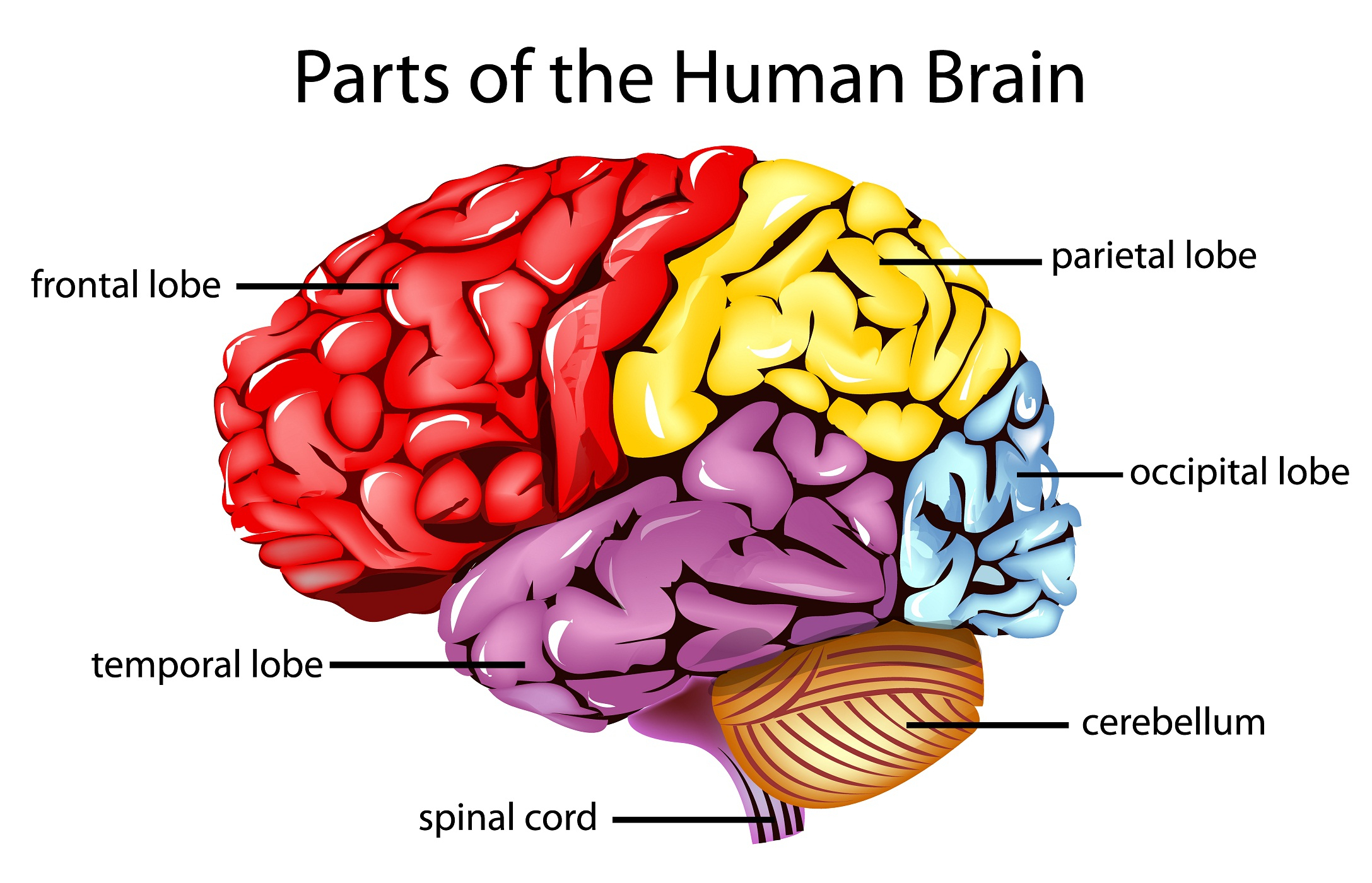
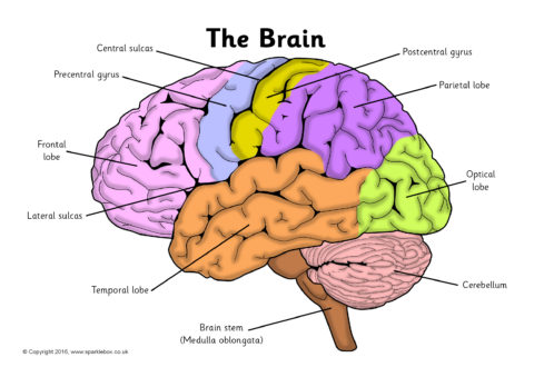
Parts of the Brain Activity for Kids, Brain Diagram, and Worksheets for ... PARIETAL LOBES - The parietal lobe provides sensory information to the brain including touch, pain and temperature. OCCIPITAL LOBES - The occipital lobe processes and interprets everything we see TEMPORAL LOBES - The temporal lobe controls emotions and short-term memory e-Anatomy: radiologic anatomy atlas of the human body - IMAIOS e-Anatomy is an award-winning interactive atlas of human anatomy. It is the most complete reference of human anatomy available on web, iPad, iPhone and android devices. Explore over 6,700 anatomic structures and more than 870,000 translated medical labels. Images in: CT, MRI, Radiographs, Anatomic diagrams and nuclear images. Available in 12 ...
75,682 Brain Anatomy Stock Photos and Images - 123RF Brain Anatomy Stock Photos And Images 75,682 matches Page of 757 Brain lobes vector illustration. Human brain infographic vector. Brain lobes functions Serotonin pathway. Humans brain with serotonin pathways. psychiatric and neurological disorders. 3D render of a medical image showing male figure with brain tumour Neocortex vector illustration.

Brain pictures and labels
PDF Automatic labeling of MR brain images through extensible learning and ... Key words: atlas selection, brain MR images, image segmentation, learning, random forest 1. INTRODUCTION Accurate brain anatomy labeling is a crucial prerequisite for numerous clinical and research applications. However, man-ual labeling is a time-consuming task because labeling a set of MR brain image requires a specialist to work for 2 or Illustration Picture of Brain Anatomy - Brain - eMedicineHealth Medical Illustrations Picture of Brain The brain is the complex organ responsible for processing sensory information (sound, touch, taste, sight, and smell). The brain controls voluntary and involuntary movements. Signals from the brain tell muscles to contract. Input from the brain controls the function of other organs in the body. Human Brain Stock Photos, Pictures & Royalty-Free Images - iStock Browse 228,812 human brain stock photos and images available, or search for human brain anatomy or human brain illustration to find more great stock photos and pictures. Experience - thin line vector icon set. Pixel perfect. Editable... Experience - thin line vector icon set. 20 linear icon. Pixel perfect.
Brain pictures and labels. Brain Label (Remote) - The Biology Corner This brain labeling activity was created for remote learners as an alternative to the labeling and coloring worksheet we would traditionally do in class. Instead of coloring and labeling on printouts, students use google slides to drag labels to the images or type the answers into text boxes. Diagram Of Brain with their Labelings and Detailed Explanation A well-labelled diagram of a human brain is given below for further reference. Structure And Function Of The Human Brain Parts Of The Human Brain The human brain is divided into three main parts: Forebrain. Midbrain. Hindbrain. These three main parts comprises many small parts. Forebrain The forebrain is also called as Prosencephalon. Labeled Brain Model Diagram | Science Trends The cerebrum is the largest and most complex portion of the human brain. The cerebrum's function is to control our actions and thoughts, either conscious or unconscious, and responses to stimuli. The cerebrum itself is typically divided into four different lobes: the temporal lobe, the parietal lobe, the occipital lobe, and the frontal lobe. Brain & Neuron Coloring Pages | Brain neurons, Neurons ... - Pinterest There are three different BIG foldables of the brain (each made of two letter pages taped together). One shows internal brain structures, another shows the lobes of the brain and the third shows the important areas of the cortex. This large format provides: 1) enough room to record descriptions of all the functions of the parts 2) three clear ...
Human Brain Anatomy - Components of Human Brain with Images ii. Cerebellum—the Sub-Cerebral Region: Placed under the cerebrum, it is a relatively smaller component of brain. Cerebellum is assigned the task of controlling and coordinating the movement of muscles, and the maintenance of balance and body posture. iii. Brainstem: Medulla + Pons + Midbrain. YouTube. Labeled brain anatomy Images, Stock Photos & Vectors - Shutterstock Labeled brain anatomy royalty-free images 2,779 labeled brain anatomy stock photos, vectors, and illustrations are available royalty-free. See labeled brain anatomy stock video clips Image type Orientation People Artists More Sort by Healthcare and Medical Anatomy Icons and Graphics human brain brain organ medicine cerebral cortex cerebellum precuneus: a review of its functional anatomy and behavioural ... Table 1 summarizes the different labels proposed for the precuneate cortex, ... such as lists of words or sets of pictures. However, memories for stimuli studied in a laboratory setting are dissimilar in important ways from naturally acquired autobiographical memories, since latter are more likely to involve complex, multimodal and emotionally salient memories embedded in a … Haloperidol Oral: Uses, Side Effects, Interactions, Pictures Find patient medical information for haloperidol oral on WebMD including its uses, side effects and safety, interactions, pictures, warnings and user ratings.
Brain Label | Human anatomy and physiology, Basic anatomy and ... Jan 26, 2014 - Image of the brain showing its major features for students to practice labeling. Answers are included. Pinterest. Today. Explore. ... Brain Label. Image of the brain showing its major features for students to practice labeling. Answers are included. Biologycorner. 17k followers . Basic Anatomy And Physiology ... Functional network organization of the human brain - PMC 17.11.2011 · The brain is a complex network with macroscopic organization at the level of functional areas and subcortical nuclei, but the number and locations of these entities in humans is largely unknown. Standard approaches to forming whole-brain rs-fcMRI graphs often ignore this issue and define nodes as voxels (e.g., Buckner et al., 2009; Cole et al., 2010; Fransson et … Labeled Diagrams of the Human Brain You'll Want to Copy Now Labeled Diagrams of the Human Brain Central Core The central core consists of the thalamus, pons, cerebellum, reticular formation and medulla. These five regions are the central areas that regulate breathing, pulse, arousal, balance, sleep and early stages of processing sensory information. Labeling Brain Structures - John Muschelli 1 Labels in template space. In Processing Within-Visit MRI, we registered the T1 image to the Eve template using a non-linear registration (SyN) (Avants et al. 2008). Also, we applied this transformation to the intensity-normalized T1, T2, and FLAIR images, so that these image are located in the same space as the Eve atlases. We can overlay the ...
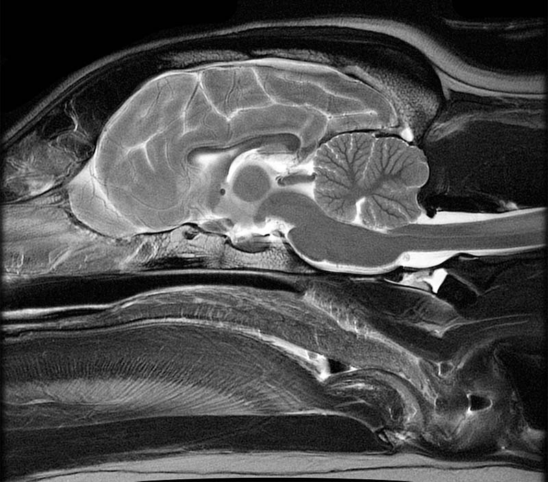
Sagittal Plane MRI Head Atlass
Split-Brain: What We Know Now and Why This is Important … 12.05.2020 · For instance, Pinto et al., 2017a) found that the split-brain patient was much better at matching pictures to sample stimuli in the left visual field. Yet, for the exact same stimuli matching pictures to verbal labels was vastly superior when the stimuli appeared in the right visual field. Crucially, response type did not play any role. The patient was better in matching …
Brain Facts & Fun - Metrowest Neurofeedback
1,000+ of the Best Brain Pictures for Free [HD] - Pixabay 1,000+ of the Best Brain Pictures for Free [HD] 1,000 Pictures of Brain in HD Related Images: people human nervous system mind Pick the perfect brain picture for your project. HD to 4K quality, available for free on all devices!

TDP-43 mutant transgenic mice develop features of ALS and frontotemporal lobar degeneration | PNAS
Human Brain Photos and Premium High Res Pictures - Getty Images 27,757 Human Brain Premium High Res Photos Browse 27,757 human brain stock photos and images available, or search for human brain anatomy or human brain illustration to find more great stock photos and pictures. Related searches: human brain anatomy human brain illustration human brain diagram human brain scan human brain vector of 100 NEXT

Sometimes, crying is the only way your eyes speak when your mouth can't explain how broken your ...
Human Brain Diagram Photos and Premium High Res Pictures - Getty Images 1,015 Human Brain Diagram Premium High Res Photos Browse 1,015 human brain diagram stock photos and images available, or start a new search to explore more stock photos and images. of 17 NEXT
Brainfeeder MP3 & Music Downloads at Juno Download
Human Brain Diagrams and Detailed Information - Innerbody The brain needs to store many different types of information that it receives from the senses and that it develops through thinking in the association areas. Information in the brain is stored in a few different ways depending on its source and how long it is needed. Our brain maintains short-term memory to keep track of the tasks in which the ...
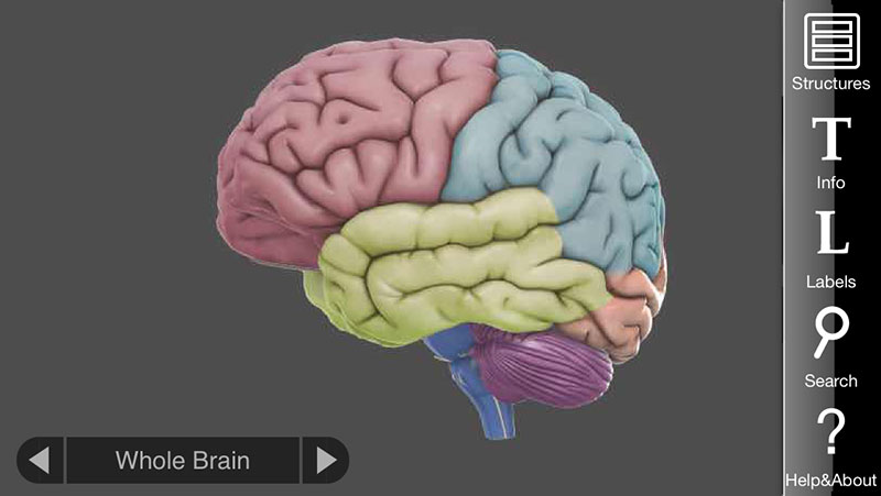
3D Brain App, an interactive way to learn about the different parts of the human brain | Apps ...
Drawing Of The Brain With Labels - Painting Valley We collected 36+ Drawing Of The Brain With Labels paintings in our online museum of paintings - PaintingValley.com. ADVERTISEMENT LIMITED OFFER: Get 10 free Shutterstock images - PICK10FREE brain human diagram labeled anatomy label system easy physiology infant coronal neat nervous spinal simple cord rat Brain Diagram Labele... 633x512 41 0

Labeling the Brain Quiz
Papers | Nicholas Carlini FixMatch first generates pseudo-labels using the model’s predictions on weaklyaugmented unlabeled images. For a given image, the pseudo-label is only retained if the model produces a high-confidence prediction. The model is then trained to predict the pseudo-label when fed a strongly-augmented version of the same image. Despite its simplicity, we show that FixMatch …
Brain Basement: Labels...good, bad, lame or excellent? by BRAIN BASEMENT
Frontiers | 101 Labeled Brain Images and a Consistent Human Cortical ... We selected 101 T 1-weighted brain MR images that are: (1) publicly accessible with a non-restrictive license, (2) from healthy participants, (3) of high quality to ensure good surface reconstruction, and (4) part of a multi-modal acquisition ( T 2*-weighted, diffusion-weighted scans, etc.).
35 Label Of The Brain - Labels Database 2020
3D Brain Image Gallery - BrainHQ from Posit Science 3D Brain Image Gallery A group of Harvard scientists has developed new methods of magnetic resonance imaging (MRI) to look more closely inside the human brain. The resulting images are stunning!
What Those Misleading Food Labels Actually Mean
Amazon.com: brain model labeled VEVOR Human Brain Model Anatomy 4-Part Model of Brain w/Labels & Display Base Color-Coded Life Size Human Brain Anatomical Model Brain Teaching Human Brain for Science Classroom Study Display Model. 3.4 out of 5 stars 3. $159.19 $ 159. 19. Get it Wed, Mar 30 - Mon, Apr 4. FREE Shipping.

Bissected Sheep Heart Matching
Brain: Atlas of human anatomy with MRI - e-Anatomy - IMAIOS Anatomy of the brain (MRI) - cross-sectional atlas of human anatomy. The module on the anatomy of the brain based on MRI with axial slices was redesigned, having received multiple requests from users for coronal and sagittal slices. The elaboration of this new module, its labeling of more than 524 structures on 379 MRI images in three different ...
Labels · william4213/Microsoft-AZ-204-Braindumps-2022 · GitHub
Parts of the brain: Learn with diagrams and quizzes - Kenhub Labeled brain diagram First up, have a look at the labeled brain structures on the image below. Try to memorize the name and location of each structure, then proceed to test yourself with the blank brain diagram provided below. Labeled diagram showing the main parts of the brain Blank brain diagram (free download!)
Label the Brain Worksheets (SB11585) - SparkleBox
Brain: Anatomy, Pictures, Functions, and Conditions The Brain Stem. PIXOLOGICSTUDIO/SCIENCE PHOTO LIBRARY / Getty Images. The brainstem is an area located at the base of the brain that contains structures vital for involuntary functions such as the heartbeat and breathing. The brain stem is comprised of the midbrain, pons, and medulla. 3.

6 nutrients that improve the well-being of mitochondria—the cell's pow | New Hope Network
Classroom Interventions for Students with Traumatic Brain Injuries 25.07.2008 · Other external cues used to remind students include labels, maps checklists, pictures or icons, photograph cues, post-it-notes, calendars, planners, and journals. A memory notebook is one such compensatory aid that has been used to assist in memory and organization following TBI. The memory notebook can be very flexible and may contain maps, checklists, …
![Gunplanerd: [CUSTOM] Bandai HG 1/550 Gipsy Avenger Full Weapon (Final Battle Ver.)](https://blogger.googleusercontent.com/img/b/R29vZ2xl/AVvXsEgLQtxrxiE2848eq1BiSIhUcKg0VYckxQWp7LHklaZdggN4r8NlZCAXufFGq38rDZWnxNyTHSHVw9pC2npWEQeR53z6b4ay2nMAluDHw9pSw_-DJtJY9OtgeYULwluZgpkgwcgZ4grLlnl3/s1600/IMG_20190406_231017.jpg)
Gunplanerd: [CUSTOM] Bandai HG 1/550 Gipsy Avenger Full Weapon (Final Battle Ver.)
101 Labeled Brain Images and a Consistent Human Cortical Labeling ... We selected 101 T 1-weighted brain MR images that are: (1) publicly accessible with a non-restrictive license, (2) from healthy participants, (3) of high quality to ensure good surface reconstruction, and (4) part of a multi-modal acquisition ( T 2*-weighted, diffusion-weighted scans, etc.).

Label parts of brain - Printable
101 labeled brain images and a consistent human cortical ... - PubMed 101 labeled brain images and a consistent human cortical labeling protocol Abstract We introduce the Mindboggle-101 dataset, the largest and most complete set of free, publicly accessible, manually labeled human brain images.

brain labeling game mc ch13 fig01 - Made By Creative Label
Neuriva Plus Brain Performance Oral: Uses, Side Effects ... - WebMD Find patient medical information for Neuriva Plus Brain Performance oral on WebMD including its uses, side effects and safety, interactions, pictures, warnings and user ratings.

31 Label Of The Brain
Assistive Technology for Individuals with Traumatic Brain Injury 11.04.2011 · Traumatic brain injury (TBI) is one of the leading causes of disability among children and young adults in the United States. According to the Centers for Disease Control and Prevention, each year an estimated 1.5 million Americans sustain a TBI. While a TBI may result in cognitive, emotional, sensory, and motor impairments, this bulletin will address assistive …
Brain Anatomy Quiz Label
Excite Hier sollte eine Beschreibung angezeigt werden, diese Seite lässt dies jedoch nicht zu.

Label the Brain Worksheets (SB11585) - SparkleBox | Human brain diagram, Brain diagram, Brain models
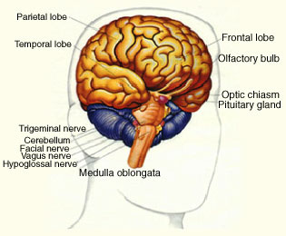
Picture of the Brain - WebMD • The cerebellum is at the base and the back of the brain. The cerebellum is responsible for coordination and balance. The brain is also divided into several lobes: • The frontal lobes are...
Post a Comment for "44 brain pictures and labels"